
Understanding Pregnancy Ultrasound: Your Baby’s First Photos
One of the most exciting moments in pregnancy is seeing your baby on an ultrasound screen for the first time. That tiny flicker of a heartbeat, those little fingers and toes, the profile of your baby’s face – these images create an emotional connection that words can’t describe. But ultrasounds are more than just photo opportunities; they’re essential medical tools that help us monitor your baby’s health and development.
As an obstetrician with over 30 years of experience, I’ve performed thousands of ultrasounds and witnessed countless magical moments. Let me guide you through everything you need to know about pregnancy ultrasounds.
What is an Ultrasound?
Ultrasound, also called sonography, uses high-frequency sound waves to create images of your baby inside the womb. These sound waves bounce off your baby’s tissues, organs, and bones, creating a picture on the screen. It’s the same technology submarines use to navigate underwater!
The procedure is:
● Completely painless
● Non-invasive
● Safe for both mother and baby
● Takes 20-45 minutes depending on the type
How Does Ultrasound Work?
During an ultrasound:
- You’ll lie on an examination table
- Gel is applied to your abdomen (it helps conduct sound waves)
- A transducer (handheld device) is moved across your belly
- Sound waves create real-time images on a monitor
- We take measurements and photos
- You get to see your baby moving!
For early pregnancy, we might use transvaginal ultrasound (inserting a probe into the vagina) for clearer images when the baby is very small.
Types of Pregnancy Ultrasounds:
1. Dating Scan (6-12 Weeks)
Purpose:
● Confirm pregnancy
● Determine how many babies you’re carrying
● Calculate due date accurately
● Detect heartbeat (usually visible around 6-7 weeks)
● Check if pregnancy is in the uterus (rule out ectopic pregnancy)
● Estimate gestational age
What We See:
At 6 weeks, your baby is tiny – about the size of a grain of rice! We can see the gestational sac and often the yolk sac. The most thrilling moment is seeing that first heartbeat flicker on the screen.
By 8-12 weeks, your baby looks more recognizable with a visible head, body, and limb buds. The heartbeat is strong and regular (120-160 beats per minute).
Why It Matters:
Accurate dating is crucial for scheduling all future tests and determining your due date. This early scan also reassures you that your pregnancy is progressing normally.
2. Nuchal Translucency Scan (11-13 Weeks)
Purpose:
● Screen for chromosomal abnormalities (Down syndrome, Edwards syndrome, Patau syndrome)
● Measure fluid at the back of baby’s neck
● Check baby’s nasal bone
● Assess heart structure
What We Measure:
The nuchal translucency is a fluid-filled space at the back of the baby’s neck. Increased thickness can indicate higher risk of chromosomal conditions. Combined with blood tests, this gives us valuable information about your baby’s health.
What You’ll See:
Your baby is now about 2-3 inches long. You can see the head, body, arms, and legs clearly. Your baby might be moving around, though you can’t feel it yet!
Important Note:
This is a screening test, not a diagnostic test. Abnormal results don’t mean something is definitely wrong – they indicate the need for further testing like amniocentesis or NIPT (Non-Invasive Prenatal Testing).
3. Anomaly Scan / Level 2 Ultrasound (18-22 Weeks)
Purpose:
This is the most detailed ultrasound of your pregnancy. We examine every part of your baby’s anatomy to check for structural abnormalities.
Comprehensive Examination:
● Brain and skull: Checking for proper development
● Face: Looking at eyes, nose, lips (detecting cleft lip/palate)
● Spine: Ensuring vertebrae are properly formed
● Heart: Examining all four chambers and major vessels
● Stomach and bowels: Confirming proper development
● Kidneys and bladder: Checking urinary system
● Arms and legs: Counting fingers and toes, checking bone length
● Placenta position: Ensuring it’s not blocking the cervix
● Amniotic fluid: Measuring fluid levels
● Umbilical cord: Checking blood flow
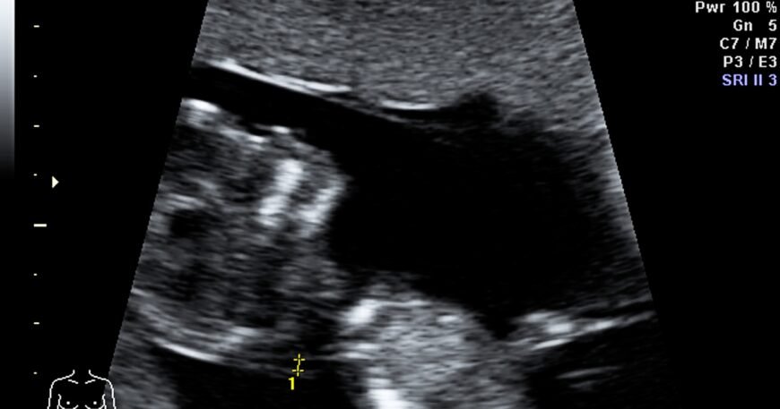
What You’ll See:
Your baby is now 6-7 inches long and weighs about 10-12 ounces. This is when ultrasounds get really exciting! You can see:
● Baby’s profile clearly
● Fingers and toes
● Baby sucking thumb
● Baby moving, kicking, and stretching
● Facial expressions
4. Growth Scans (Third Trimester)
Purpose:
Not all women need growth scans, but we recommend them if:
● Baby seems small or large for gestational age
● You have gestational diabetes
● You have high blood pressure
● You’re carrying multiples
● There are concerns about placental function
● Previous pregnancy had growth issues
● Decreased fetal movements
What We Measure:
● Head circumference
● Abdominal circumference
● Femur length (thigh bone)
● Estimated fetal weight
● Amniotic fluid volume
● Placental health
● Blood flow through umbilical cord (Doppler study)
These measurements help us determine if your baby is growing appropriately.
5. Biophysical Profile (BPP)
When Needed:
Usually performed in late pregnancy if there are concerns about baby’s well-being, especially in high-risk pregnancies or post-dates pregnancies.
What We Check:
● Fetal breathing movements
● Fetal movements
● Fetal tone (flexion and extension)
● Amniotic fluid volume
● Non-stress test (monitoring heart rate)
Each parameter is scored, giving us an overall picture of baby’s health.
6. 3D and 4D Ultrasounds
What’s the Difference?
● 2D: Traditional flat, black-and-white images
● 3D: Three-dimensional still images showing baby’s features
● 4D: Live 3D images showing baby moving in real-time
Purpose:
Primarily for bonding and keepsake photos, though 3D can sometimes help diagnose certain facial abnormalities like cleft lip.
Best Timing:
26-30 weeks is ideal for 3D/4D ultrasounds. Baby is developed enough to see features clearly but not too cramped yet.
What to Expect:
You’ll see your baby’s face, expressions, possibly yawning or sucking thumb. It’s an incredible bonding experience! However, good images depend on:
● Baby’s position
● Amount of amniotic fluid
● Your body composition
● Baby’s cooperation!
What Doctors Look for During Ultrasounds:
First Trimester:
● Confirmation of intrauterine pregnancy
● Heartbeat presence and rate
● Number of embryos
● Gestational age
● Early anatomical development
Second Trimester:
● Detailed anatomy survey
● Markers for chromosomal abnormalities
● Placental location
● Cervical length
● Growth parameters
Third Trimester:
● Fetal growth and weight
● Position (head down vs. breech)
● Placental function and location
● Amniotic fluid levels
● Blood flow patterns
● Baby’s well-being
Common Ultrasound Findings:
Normal Variations:
● Choroid plexus cysts (usually resolve by 26 weeks)
● Single umbilical artery (usually not significant)
● Echogenic bowel (may need follow-up)
● Minor heart variations
Conditions We Might Detect:
● Placenta previa (placenta covering cervix)
● Low amniotic fluid (oligohydramnios)
● Excess amniotic fluid (polyhydramnios)
● Growth restriction
● Macrosomia (large baby)
● Structural abnormalities
● Multiple pregnancy complications
Is Ultrasound Safe?
Absolutely! This is one of the most common concerns I hear.
The Facts:
● Ultrasound has been used for over 40 years
● Extensive research shows no harmful effects
● No radiation involved (unlike X-rays)
● Professional medical organizations worldwide endorse its safety
● Billions of babies have been safely monitored with ultrasound
Best Practices:
● Ultrasounds should be performed by trained professionals
● Use the lowest power necessary for adequate images
● Minimize scan duration while getting needed information
● Medical ultrasounds only (recreational frequent scans not recommended)
Preparing for Your Ultrasound:
Before the Appointment:
● Early pregnancy: You may need a full bladder (helps visualize baby better)
● Later pregnancy: Usually no preparation needed
● Wear comfortable, loose clothing
● Bring your partner or support person
● Bring tissues (for gel cleanup and happy tears!)
During the Scan:
● Try to relax (stress can make baby move away from the transducer)
● Ask questions – we’re happy to explain what you’re seeing
● Don’t be disappointed if baby isn’t cooperative (sometimes they face away or have hands in front of face)
● Remember that medical assessment comes first, photo opportunities second
Understanding Your Ultrasound Report:
Your report will include:
● Gestational age: Based on measurements
● EDD: Estimated due date
● Presentation: Baby’s position (head down, breech, transverse)
● Placental location: Where placenta is attached
● AFI: Amniotic Fluid Index (normal is 5-25 cm)
● EFW: Estimated fetal weight
● Biometry: Measurements of head, abdomen, femur
● Anatomical survey: Checklist of examined structures
When Additional Ultrasounds Are Needed:
Medical Indications:
● Vaginal bleeding
● Decreased fetal movement
● Measuring too small or large
● High blood pressure
● Diabetes
● Previous pregnancy complications
● Multiple pregnancy
● Suspected placental problems
● Post-dates pregnancy (after 40 weeks)
What If Something Abnormal is Found:
If we detect an abnormality:
- Stay calm: Many issues can be managed successfully
- Get more information: We may recommend additional testing or specialist consultation
- Understand the significance: I’ll explain what the finding means for your baby
- Discuss options: We’ll talk about management and treatment options
- Get support: You’re not alone – we’re here to support you
Special Situations:
Twins or Multiples:
More frequent ultrasounds to monitor:
● Growth of each baby
● Chorionicity (shared or separate placentas)
● Twin-to-twin transfusion syndrome risk
● Growth discordance
Breech Baby:
Ultrasound helps determine:
● Type of breech position
● Baby’s size
● Placental location
● Whether external cephalic version (turning baby) is safe
Previous C-Section:
Check placental location to rule out:
● Placenta previa
● Placenta accreta (abnormally attached placenta)
The Emotional Aspect:
Ultrasounds are powerful emotional experiences:
● Seeing your baby makes pregnancy feel real
● Hearing the heartbeat is incredibly reassuring
● Watching your baby move creates a bond
● Finding out the gender is exciting
● Detecting problems can be devastating
Remember: I’m here not just as your doctor but as your support through this journey.
Common Questions:
Q: How many ultrasounds will I have?
A: Typically 2-3 for low-risk pregnancies:
● Dating scan (first trimester)
● Anomaly scan (20 weeks)
● Growth scan if needed (third trimester)
High-risk pregnancies may need more frequent monitoring.
Q: Can ultrasound harm my baby?
A: No. Decades of research and billions of safe ultrasounds confirm that diagnostic ultrasound doesn’t harm babies when performed by trained professionals.
Q: Why can’t you always see the gender?
A: Sometimes baby’s position makes it impossible to see, or they might have legs crossed. We never guess – we’ll tell you if we can’t see clearly.
Q: Should I get 3D/4D ultrasounds at commercial studios?
A: While not harmful, I recommend medical ultrasounds be performed in medical facilities by trained professionals. Commercial “keepsake” ultrasounds shouldn’t replace medical monitoring.
Q: What if baby isn’t measuring right on dates?
A: Small variations are normal. We look at growth trends over time, not just single measurements. If there are concerns, we’ll monitor more closely.
Q: Can you see everything on ultrasound?
A: Ultrasound is excellent but not perfect. Some conditions can’t be seen on ultrasound, and some only become apparent later in pregnancy or after birth.
My Approach to Ultrasounds:
At our clinic:
● We use state-of-the-art ultrasound equipment
● I personally perform most ultrasounds
● We take time to show you your baby and explain what we’re seeing
● You’ll get printouts to take home
● We create a comfortable, private environment
● We’re sensitive to your emotional state
● We provide clear explanations of all findings
The Bottom Line:
Pregnancy ultrasounds are one of modern medicine’s greatest gifts. They allow us to monitor your baby’s development, detect potential problems early, and give you precious glimpses of your little one before birth. While the medical information is crucial, never underestimate the emotional and psychological benefits of seeing your baby growing healthy and strong.
Those grainy black-and-white images might not look like much to others, but to you, they’re the first pictures of your child – a preview of the love story that’s about to unfold.
Schedule Your Ultrasound:
Whether you need a routine dating scan, detailed anomaly scan, or follow-up growth scan, we’re here to provide expert ultrasound services in a warm, caring environment. Call +91-9811078323 or message us on WhatsApp. Walk-ins welcome Monday-Saturday, 10 AM – 7 PM.
- Dr. Deepa Dureja
MD (Obstetrics & Gynaecology)
30+ Years Experience
S-77, Greater Kailash Part 2, South Delhi